Necrosis of the hard tissues of the tooth
Machine translation
Original article is written in RU language (link to read it) .
Tooth tissue necrosis is a severe pathology that often leads to tooth loss.
Glass ionomer cement (GIC) and resin-modified glass ionomer cement (RMGIC): advantages, indications, and clinical application in the webinar New materials in pediatric dentistry.
The disease can be caused by various external and internal causative factors. The endogenous ones include the following:
- disorders of the functioning of the endocrine glands,
- central nervous system disorders,
- hereditary tooth diseases,
- chronic intoxications of the patient.
Cervical necrosis
A type of this non-carious tooth pathology is cervical necrosis – a pathology characteristic of patients with hyperthyroidism, pregnant women. The highest intensity of the disease is observed when hyperthyroidism is combined with pregnancy.

Figure 1. Enamel defect in the cervical area.
Severe symptoms of thyrotoxicosis include disturbances in mineral and protein metabolism. Typical clinical manifestations include:
- formation of necrosis areas on the vestibular surface primarily in the region of the necks of the frontal teeth, less often - molars and premolars;
- initially, minor chalky streaks are diagnosed in the cervical zone, the surface of which is shiny and smooth;
- gradually, the area of affected enamel fragments increases, the surface loses its shine, becomes rough, and the enamel has a matte appearance;
- over time, the enamel on the affected area completely disappears, exposing the dentin;
- the boundaries of the defect are unclear, with a tendency to increase;
- in some patients, against the background of a lack of thorough hygiene, a carious cavity forms in the area of the defect;
- the pathological process gradually spreads over the entire surface;
- the enamel is so loose that it can be scraped off with an excavator without effort;
- cervical necrosis, especially at the stage of loss of the enamel layer, is in most cases accompanied by hyperesthesia.
A patient diagnosed with cervical necrosis must undergo an examination by an endocrinologist. For high hypersensitivity of the necks, agents that help reduce its intensity or eliminate it are used. If dentin damage is observed and a carious cavity has formed in the area of the necrotic focus, tooth filling is necessary. It is only important to consider the fact that in the future, the enamel around the restoration may again undergo necrosis; before filling, it is advisable to conduct remineralizing therapy, which will help strengthen the tooth tissues.
Acidic Necrosis
Chemical, or acidic, necrosis is the result of local exposure. This pathology is observed in patients who have worked for a long time in the production of inorganic or organic acids. In the absence of quality ventilation in the air of these production workshops, acid vapors accumulate, which, when entering the mouth, remain in the saliva. The latter acquires an acidic reaction, which subsequently causes decalcification of the tooth tissues.
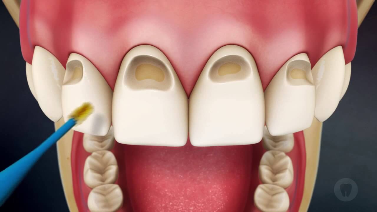
Figure 2. Closure of defects in the cervical area.
Initial manifestations of acid necrosis include the onset of numbness and a feeling of disgust in the teeth. Sometimes the impact of chemical and temperature irritants is accompanied by pain, and there may be a sensation of teeth sticking together during occlusion. Gradually, these sensations disappear or become dull due to the formation of reparative dentin, the development of dystrophic changes or necrosis in the pulp.
The enamel of the front teeth during an objective examination is matte, rough, and dirty-gray in shade. The wear of the teeth is significantly pronounced. Along the cutting edge, the enamel disappears, forming sharpened, easily breakable fragments of the crown, and then the process of wear and destruction affects the tooth tissues not only on the vestibular but also on the lingual surface of the frontal group. Over time, the crowns of the front teeth are destroyed down to the level of the gums, and the premolars and molars are heavily worn.
Prevention of chemical necrosis primarily involves ensuring proper supply and exhaust ventilation to seal the production process. In the workshops, devices with alkaline water are placed where employees must rinse their oral cavity. In treating acid necrosis, it is necessary to eliminate the influence of the acidic agent, subject the patient to remineralizing therapy, and then proceed with filling using glass ionomer cements.
Toxic Necrosis
Develops as a result of frequently inhaling vapors of acetone, gasoline, and other similar substances. It occurs more often in teenagers.
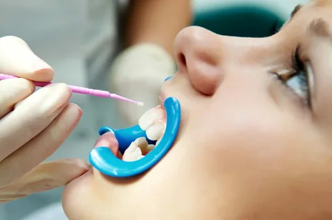
Figure 3. Enamel remineralization.
Typical clinical manifestations
- in the early stages, the enamel acquires a light-yellow or dark-brown color,
- the enamel loses its shine, the surface becomes rough;
- areas of demineralization appear;
- over time, extensive necrosis fragments form, their surface covered with loose necrotic masses.
Patients in this group require regular medical monitoring.
Post-radiation, or radiation, necrosis
Develops as a result of the influence of ionizing radiation during the treatment of malignant tumors, as well as under the influence of harmful production factors.
The pathogenesis of radiation necrosis has not been fully established. The disease has a chronic course. Pathology of tissue respiration in its aerobic phase is accompanied by the accumulation in tissues, including dental pulp, of under-oxidized metabolic products, typically causing persistent disruption of their subsequent oxidation to carbon dioxide and water. Under the influence of ionizing radiation, these processes occurring in the dental pulp cause disruption of trophism and natural mechanisms of tissue remineralization of the tooth.
Clinical manifestations of post-radiation necrosis:
- radiomucositis of the mucous membranes of the oral cavity,
- distortion or loss of taste sensitivity,
- xerostomia,
- acute caries,
- radiation necrosis.
The foci of necrosis are initially local, then they spread circularly across the tooth surface, acquire a dark color, are painless, and are filled with soft necrotic masses. Occlusal and buccal surfaces of the teeth are brittle and worn.
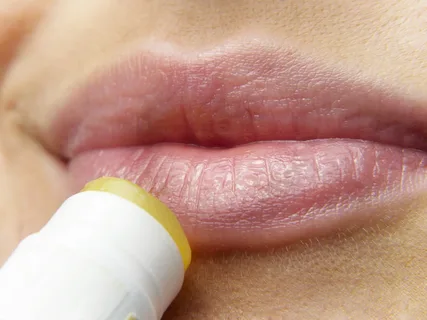
Figure 4. Dry lips.
Characteristics of acute caries in post-radiation necrosis:
- the edges of carious cavities are uneven, eroded,
- cavities are filled with loose dirty-gray masses, which are removed almost painlessly;
- fillings fall out, both old and newly placed;
- electroodontometry indicators are reduced.
Prevention of radiation necrosis involves minimizing direct radiation exposure, which is achieved by using a lead cap placed in the patient's mouth before each session of radiation therapy. It is important to reduce the direct impact of penetrating radiation through a preliminary (the day before radiation) course of remineralizing therapy combined with antioxidants.
Sealing is possible after a month to a month and a half with the mandatory preliminary use of a therapeutic pad based on calcium hydroxide, which has an odontotropic effect. Teeth are sealed with glass ionomer cement exclusively for this pathology.
Computer Necrosis
Clinical manifestations of computer necrosis are characterized by multiplicity, systematic nature, and extensive damage to the tooth. Necrosis areas occupy a significant, sometimes large part of the tooth surface, including caries-resistant areas. These lesions are predominantly pigmented: dark brown, almost black in color, represented by loose masses of dirty brown shade, which are easily removed using an excavator, but are painful. Unaffected fragments are grayish-white or cloudy-white in color, lacking luster. Mild hyperesthesia is characteristic of the initial pathological process.
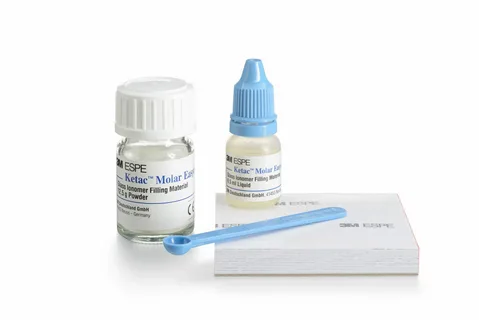
Figure 5. Glass ionomer cement for filling.
All patients report hyposalivation, which gradually transitions into xerostomia. The roots of the teeth are exposed, especially from the buccal side, with resorption of the alveolar bone and interdental septa observed.
Radiological signs: the pattern of the alveolar bone and tooth is unclear, confirming the hypomineralization of these anatomical formations.
In the blood, biochemical analysis shows no deviations, but patients have slowed blood coagulation, and tooth extraction requires the mandatory use of hemostatic agents and prior preparation of the patient. Extracted teeth are brittle and often crumble in the forceps.
In the first stage, local treatment involves cleaning the tooth from necrotized tissues, conducting remineralization through daily applications of phosphate-containing agents. After a few months, selective filling of individual teeth can begin.
About the use of glass ionomer cement for creating direct restorations in the webinar Aesthetic Restorations in Pediatric Dentistry. Making the Right Clinical Decisions.