
The Importance of Diagnostics in Endodontics
Machine translation
Original article is written in RU language (link to read it) .
Diagnosis is a direction of thought, the result of analyzing subjective and objective data. Teaching professional thinking, the ability to see, hear, assess the condition of teeth and oral cavity, and perform detailed diagnostics of a specific tooth is the main task in training dentists.
Diagnostic methods for various endodontic problems are presented in the webinar Endo-periodontal lesions: diagnosis and treatment.
Direct contact with the patient, a psychosocial approach, building trusting relationships - these are the foundation of practical work in clinical dentistry. The inability to establish a dialogue with the patient is one of the leading factors contributing to errors in diagnostics, treatment planning, and an unfavorable prognosis of endodontic treatment.
Stages of Diagnosis
The first stage of diagnosis is an interview, and to avoid potential errors at this stage of the examination, it is important:
- to establish complaints, based on which a certain nosological form of the disease can be assumed;
- to identify the presence of risk factors contributing to the development of dental diseases;
- to assess the overall condition of the patient (presence of allergies, systemic diseases).
The second stage of diagnosis is an examination, correct determination of the dental status significantly improves the prognosis of endodontic treatment. Visual inspection is accompanied by an index assessment of the oral cavity condition.
Figure 1. The importance of diagnostics in endodontics.
The third stage – detailed examination of a specific tooth involves the use of diagnostic techniques (palpation, percussion, determination of EOD, radiography), and a mandatory evaluation of the tooth crown and the condition of its roots.
Establishing the correct diagnosis of a problematic tooth mainly depends on adhering to the stages of patient examination and the meaningfulness of medical diagnostic manipulations. The main difficulties in diagnosing some forms of pulpitis and apical periodontitis are associated with the asymptomatic course of the disease.
Radiological Examination
In the process of diagnosing complications of caries, a radiograph is used to detect the following problems:
- denticles,
- granulomas and cystogranulomas,
- signs of external and internal resorption,
- assess the condition of the canal, its anatomical features,
- identify pathological changes in the tissues of the periodontium.
A common cause of diagnostic errors is incorrect interpretation of radiological examination results. On the upper jaw, the incisive foramen or maxillary sinus may overlap the area of the root apex. On the lower jaw, the mental foramen is sometimes mistaken for a focus of bone destruction at the apex.
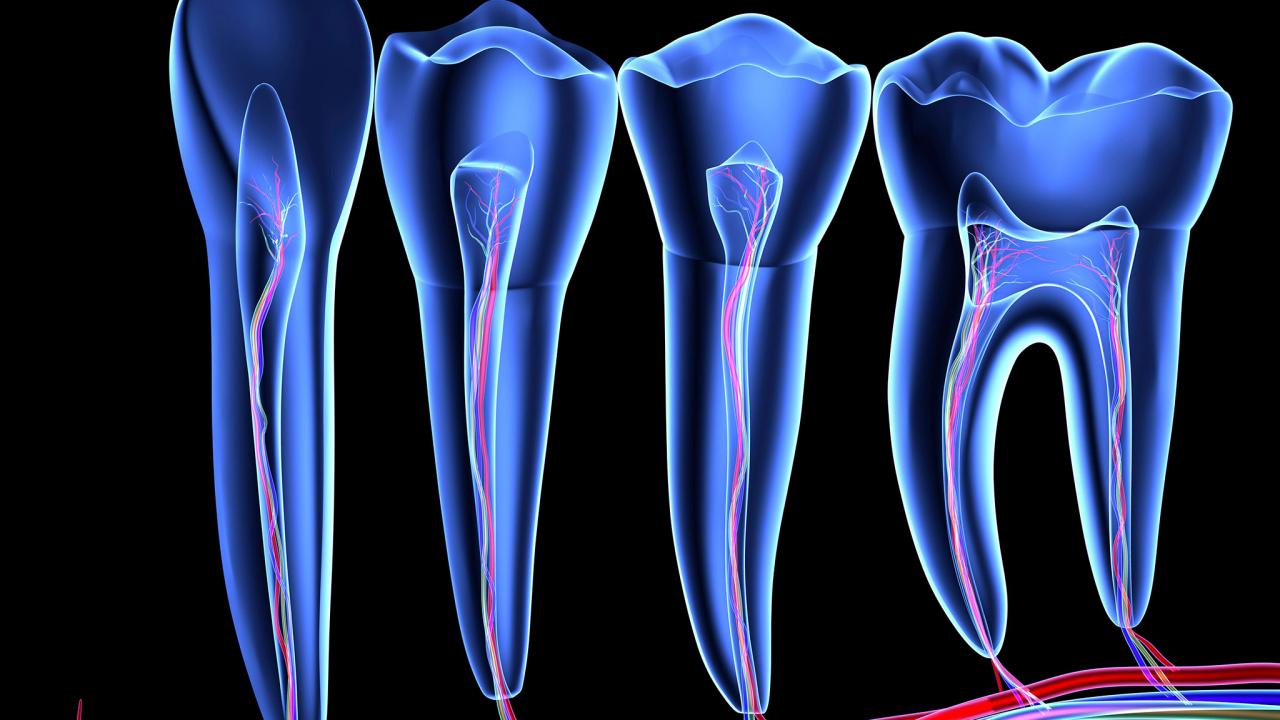
Figure 2. Topography of the pulp space in different teeth.
A reliable assessment of the condition of teeth on an X-ray during endodontic treatment is not only of diagnostic value but is also very important for the prognosis of the treatment. Often the informativeness of radiological methods is reduced due to the superimposition of several anatomical formations, foci of destruction, and root apices on the image. This is a common cause of diagnostic and subsequently tactical errors in endodontic treatment of various forms of pulpitis and apical periodontitis.
The use of CBCT significantly expands the capabilities of radiographic diagnostics, providing a three-dimensional image of anatomical structures, including a layered cross-sectional image of the root along its entire length, which is especially important for endodontic practice. The absence of radiological examination on the eve of and during endodontic treatment leads to a number of complications (perforation of the pulp chamber floor or root wall), which is due to underestimating the curvature of the canal or the topography of the bifurcation, as well as other pathological processes not detected before preparation.
During the actual endodontic treatment, the radiographic image allows monitoring the working length and the quality of the mechanical processing of the canal, assessing the quality of the filling and its long-term results.
Diagnostic Errors
Often, the occurrence of errors is due to the doctor's ability to apply diagnostic tests and analyze the collected anamnesis data. To avoid this mistake, the doctor needs to have extensive theoretical knowledge about the causes and pathogenesis of complications of the carious process, be able to analyze symptoms, and correctly interpret the results of the examination.
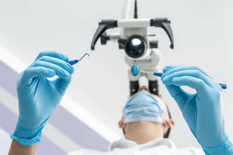
Figure 3. Microscopic endodontics.
Adhering to the technique of instrumental assessment of tooth tissues aids in accurate diagnosis. For instance, probing the cavity is necessary to check the density of tissues, detect communication with the tooth cavity, and locate the canal orifices.
Vertical percussion allows assessing the condition of the apical periodontium; this method is particularly informative in cases of pronounced symptoms characteristic of acute inflammatory processes in the apical periodontal tissues.
Diagnosing the reaction of a diseased tooth to temperature stimuli provides information about the condition of the pulp. For example, the pain response in chronic pulpitis is stronger to cold, while in the case of gangrenous or purulent processes, the tooth pulp is irritated by hot water.
An important aspect is the differential diagnosis of deep caries with irreversible pulp damage and possible involvement of the apical periodontium, which clinically proceed asymptomatically. In these situations, to clarify the diagnosis, it is recommended to conduct EOD (Electric Pulp Testing), but in practice, this technique is limited due to the frequent absence of equipment.
However, it is important to know and be able to interpret EOD readings in any clinical situation, as they significantly assist in choosing the tactics of endodontic treatment. In chronic pulpitis, the EOD value ranges from 30-45 µA; if the indicator exceeds 60 µA, it indicates the death of the pulp in the coronal part of the tooth, and a value above 100 µA indicates complete pulp necrosis (diagnosis of periodontitis).
The final stage of diagnosis is dissection, which allows for the removal of pathological tissues, determining the volume and depth of tooth structures affected by the carious process.
Diagnostic dissection involves preparation for diagnosis. This preparation includes the following stages:
- opening,
- expansion of the cavity,
- necrectomy.
It is important to consider the morphometric values of hard tissues, which help to determine the depth of the cavity formed at the end of necrectomy, and probing shows a dense bottom, the presumed diagnosis is moderate or deep caries.
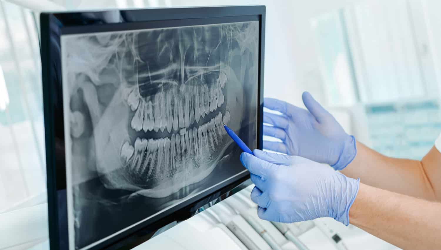
Figure 4. Radiography – an important stage in diagnostic during endodontic treatment.
To prevent errors during the diagnostic stage, it is important to remember that the main object of endodontics is the pulp space. It is not always possible to determine the condition of the pulp, it is difficult to assume how many microbes have penetrated into the pulp chamber, the depth of their penetration (the time factor of bacterial flora activity), and what changes have occurred in the pulp cavity, at what levels.
In cases where deep damage is found against the backdrop of asymptomatic disease progression, it is important to assess the condition of the pulp in the crown part (central chamber and peripheral zone), and when diagnosing partial or complete death of the crown pulp, irreversible forms of pulpitis can be assumed. If at the final stage of diagnosis any level of the canal still contains remnants of pulp that require extirpation, this indicates chronic pulpitis. Incorrect assessment of the pulp condition provokes incorrect diagnosis, errors in choosing treatment tactics, and subsequently leads to the development of a more serious complication – apical periodontitis.
For the prevention of errors in the treatment of complicated forms of caries, endodontic preparation of the pulp space using appropriate tools for its different levels is of important diagnostic value. Failure to follow the stages of crown and root preparation leads to the most serious errors in endodontic treatment.
Knowing the purpose of any stage of endodontic preparation will help minimize the risk of errors and complications.
Hence the importance of understanding the tasks and sequence of crown preparation:
- creating access to the pulp chamber,
- complete removal of the chamber roof, if necessary — pulp amputation,
- preparation of the walls and floor of the tooth cavity for a smooth transition to the canal orifices.
Mastering the knowledge of the peculiarities of endodontic preparation of teeth of different group affiliations reduces the likelihood of errors during endodontic treatment.
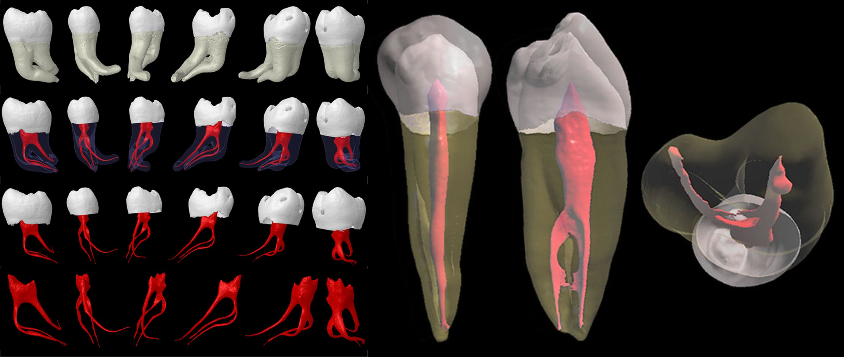
Figure 5. Anatomical and topographical features of the pulp space of different teeth.
At the stage of endodontic preparation of the root part of the tooth, errors are associated with a lack of knowledge about the features of teeth in the second projection, these features are not determined radiologically.
In endodontics, the success of the final diagnosis is determined by the clarity of the root and crown preparation stages. In this regard, it is always necessary to consider the anatomical and topographical characteristics and morphological features of each tooth, know the features of teeth in three projections, this guarantees a favorable result of endodontic treatment.
The principles of diagnostics in emergency conditions in endodontic practice are detailed in the online course Emergency Conditions in Endodontics: Diagnosis, Treatment.