
Mistakes in the Mechanical Processing of Channels
Machine translation
Original article is written in RU language (link to read it) .
During the endodontic treatment of canals, errors can occur at any stage of treatment. For example, during the preparation of canal orifices for subsequent endodontic treatment, the following errors are possible:
- Insufficient opening of the canal orifices.
- Perforation of the canal wall or the floor of the pulp chamber.
- Excessive opening of the canal orifices.
The technique for forming an apical plug is described in the webinar Working with Perforations and Wide Apical Openings.
Insufficient opening of the canal orifices subsequently causes difficulties when attempting to insert a file into the root canal, which will lead to time consumption, inevitable bending or breaking of the instrument, the likelihood of forming a false canal, and creates difficulties for the obturation of the root canal. It also leads to increased load on the files, which may break during subsequent work.
Prevention: adequate opening of the canal orifices.
Perforation of the pulp chamber floor occurs due to the doctor's lack of experience, unfamiliarity with the tooth cavity layout, and lack of precision in work. This mistake significantly worsens the prognosis of endodontic treatment and leads to additional time and material costs. If pathology from the periodontal tissues joins, this mistake requires the tooth to be mandatorily removed.
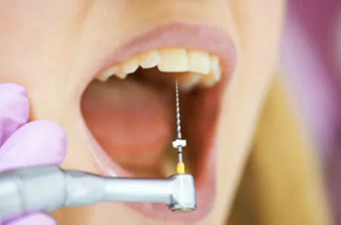
Figure 1. Endodontic treatment of a tooth.
Prevention: studying the layout of the tooth cavity, following the protocol for forming access to the canals, using optical magnification. Error correction: sealing the perforation with special materials.
Excessive opening of the canal orifices is often observed during the mechanical processing of curved canals when the sequence of file use is not followed. This error increases the likelihood of canal wall perforation during processing; complicates the insertion of the file into the canal, which significantly increases the duration of the manipulation, and also negatively affects the condition of the small-sized endodontic instruments used at the initial stage of canal passage, increasing the risk of fracture.
Preventive measures: gentle preparation in the area of the canal orifice. Remediation: smooth the ledge, if it is impossible to perform processing and subsequent filling considering its presence.
Canal perforation is a consequence of active work in the area of the orifice with a machine tool, an attempt to smooth the ledge in the orifice third, during the canal refilling.
Risk factors for canal wall perforation:
- morphological features of the tooth,
- presence of additional canals.
This error leads to the inability to properly prepare the canal for obturation, and may require surgical treatment (tooth extraction or apical resection).
Prevention: improving manual skills, mastering the technique of working at the canal orifice, using tools with non-aggressive tips, and creating a "carpet path" before starting to work with machine files.
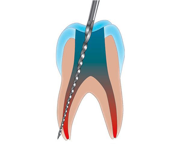
Figure 2. Perforation of the canal wall.
Error correction: sealing the perforation, thorough canal treatment, using a non-irritating sealer for the surrounding tissues.
Errors during the working length determination stage
At this stage, two types of errors are possible: not determining the working length or incorrect determination.
Neglecting the stage of determining the working length is a serious violation in endodontic treatment. Treating a canal without knowing its length can lead to either underfilling of the canal or cause the instrument to protrude and spread the infection to the periapical tissues, which further leads to the extrusion of the filling material beyond the apex.
Prevention: knowing the methods of determining the working length and strictly adhering to this stage.
Incorrect calculation of the working length
Possible reasons include:
- presence of residual EDTA gel in the canal during the electrometric method of determining the working length distorts the device readings, as EDTA has increased electrical conductivity;
- errors are not accounted for when using the radiographic method.
Prevention: thorough study and careful adherence to the methods of determining working length.
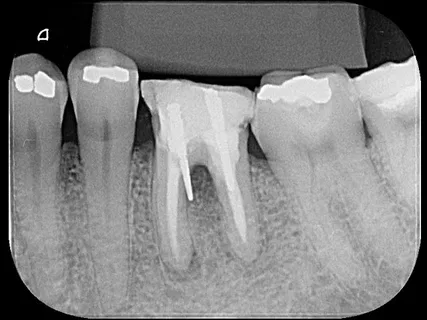
Figure 3. Radiological signs of root canal perforation.
Errors during the mechanical processing of the canal
At this stage, the following errors can be identified:
- Pushing infected tissues beyond the apex.
- Injuring the growth zone (when treating children).
- Incomplete cleaning of the canal from pulp residues.
- Inadequate mechanical processing of the canal.
- Creating a defect in the lower third.
- Extending the file beyond the apex.
- Perforation of the canal wall.
- Irritating effect of medications.
- Breakage of a part of the endodontic instrument, its jamming in the canal.
- Blocking the canal with filings.
Pushing Infected Tissue Beyond the Apex
This often occurs during rapid, careless work in the canal, or when there is a gaping apical opening in a specific canal. The consequence is additional infection of the surrounding tissues.
Prevention: meticulous adherence to work stages, carefulness, thorough removal of infected tissues under antiseptic baths.
Methods of elimination: anti-inflammatory therapy. Radiological control over the next six months for timely diagnosis of complications.
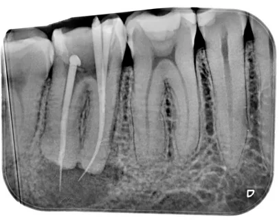
Figure 4. Result of pushing infected tissues beyond the tooth apex.
Trauma to the germinal zone
Careless work in a tooth canal with an undeveloped root can lead to disruption of the germinal zone. Prevention: radiographic control, careful adherence to working length; consideration and knowledge of root formation periods.
Incomplete cleaning of the canal from pulp residues
Possible reasons:
- incorrectly set working length;
- the canal is curved or impassable;
- not all canals were found.
Pulp residues will further provoke "residual" pulpitis, or cause inflammation in the periodontal tissues.
Preventive measures: knowledge of the number and topography of canals in a specific tooth; accurate calculation of working length; radiographic control.
Inadequate mechanical processing of the canal
Passing through the canal is just the beginning, it is important to thoroughly process it. The walls of the canals in periodontitis are covered with a microbial biofilm, traces of which are also found in the dentinal tubules. It must be removed to achieve quality obturation later.
Creating a defect in the lower third
There are two types of this error:
- Formation of a ledge – caused by the unjustified choice of a large-sized tool, non-compliance with the endodontic access protocol, canal blockage. This increases the risk of perforation; complicates canal traversal.
- Violation of the apical foramen – this error is caused by incorrect canal instrumentation technique, skipping the stage of working length determination, or an error at the determination stage. Complications: toxic effects of antiseptics on the periodontium, difficulties during obturation, possibility of material extrusion beyond the apex.
Extrusion of the file beyond the apex
Possible complications of this error include:
- traumatization of periapical tissues,
- formation of a hematoma,
- infection of intact periodontal tissues, development of inflammation,
- post-filling pain,
- increased duration of treatment,
- risk of material extrusion beyond the apex during obturation.
Canal Wall Perforation
Most commonly occurs during mechanical treatment of difficult, curved canals. Types of perforations:
- Apical, when the main canal curves in the lower third.
- Lateral – perforation in the area of the middle third on the minor curvature, caused by underestimating the morphological features of the root, curvature of the canal. Observed with significant canal enlargement, working with machine tools.
Treatment: sealing the perforation. The prognosis for tooth treatment significantly worsens.
Irritating Effects of Medications
The agents used for canal irrigation are potent substances, capable of causing local irritation, even systemic neurotoxic reactions.
Precautions: meticulous adherence to application techniques, avoid extruding the solution beyond the apex, its concentration must match specific clinical conditions.
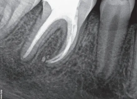
Figure 5. Extrusion of material beyond the apex.
Fracture of a part of the endodontic instrument, its getting stuck in the canal
Possible causes:
- lack of direct access to the canals,
- difficult passages,
- repeated sterilization of instruments,
- lack of a complete set of files for passing and expanding,
- non-compliance with the protocol: sequential change of instruments, refusal from EDTA, insufficient irrigation of canals, incorrect technique of using instruments.
Canal blockage by filings
Or impairment of canal patency. This error is due to not adhering to the need to return to the initial file ("recapitulation") to maintain free access to the lower third of the canal, or poor irrigation, which leads to the accumulation of dentin filings that hinder the advancement of the instrument along the canal. This complication can increase the duration of treatment or lead to incomplete processing and poor-quality obturation.
Tools and techniques for treating perforations are described in detail in the webinar Root Perforations - Diagnosis and Treatment.